[from LLNL’s Science & Technology Review, by Sheridan Hyland]
Respiratory diseases can balloon from a few cases to epidemics or pandemics, making them a consideration in national security planning and preparation. Understanding how a pathogen behaves in different circumstances and environments is complex and depends upon many factors, including genetics. COVID-19 remains a stark example of how understanding of pathogen behavior can be tenuous. Scientists continue to grapple with answering questions of why some carriers are asymptomatic while others produce symptoms of the disease, why some of the infected suffer the effects of long COVID while others do not, and why some individuals passed away and others survived. COVID-19 is only one example of a pathogen that raises questions among medical researchers: Valley Fever, variations of encephalitis, and other diseases also present additional challenges.
Lawrence Livermore scientists are taking a comprehensive approach to understanding pathogen behavior and immune system response, building on past research that focused on individual molecules and interactions. Dina Weilhammer, a staff scientist in Livermore’s Biosciences Division, explains, “We’ve made significant inroads in understanding how the immune system recognizes foreign agents such as bacteria and viruses, as well as how the immune system can separate what should be eliminated and what should be protected. Today’s biotechnology tools can overcome earlier limitations. They enable us to interrogate the immune system more holistically than ever before to address questions and uncover unexpected considerations and answers.”
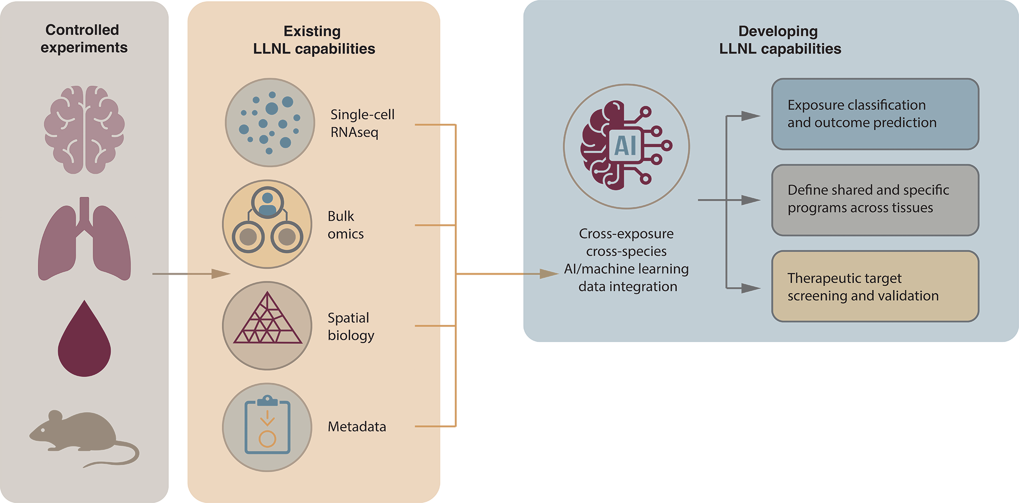
Through its research, the Livermore team—which includes Weilhammer, Nicholas Hum, Aimy Sebastian, Oscar Davalos, Margarita Rangel, Deepa Murugesh, Bria Gorman, Gregory Lum, Nicole Leon, Patrick Hurley, and Ashlee Phillips—is sharpening its understanding of the differences between one immune system that clears infection compared to another that cannot. Using a combination of controlled pathogen exposure; single-cell RNA sequencing (scRNA-seq); spatial transcriptomics to collect data; and computational platforms to organize, analyze, and interpret the data, the Laboratory is developing an immune system atlas that could help the medical field better understand and predict disease outcomes in the future.
Mapping the Methodology
To address their research questions, scientists developing the immune atlas have focused on pathogens that impact the lungs and brain. “We have viral, fungal, and bacterial models of disease that specifically target the lungs,” says Hum, a staff scientist. “At the same time, many diseases do not start in the lungs but can still affect them over time through systemic immune and inflammatory responses. The next natural question is how signals from elsewhere in the body reshape the lung environment and influence lung function.” Data from multiple techniques combine to provide a fuller picture of immune system response.
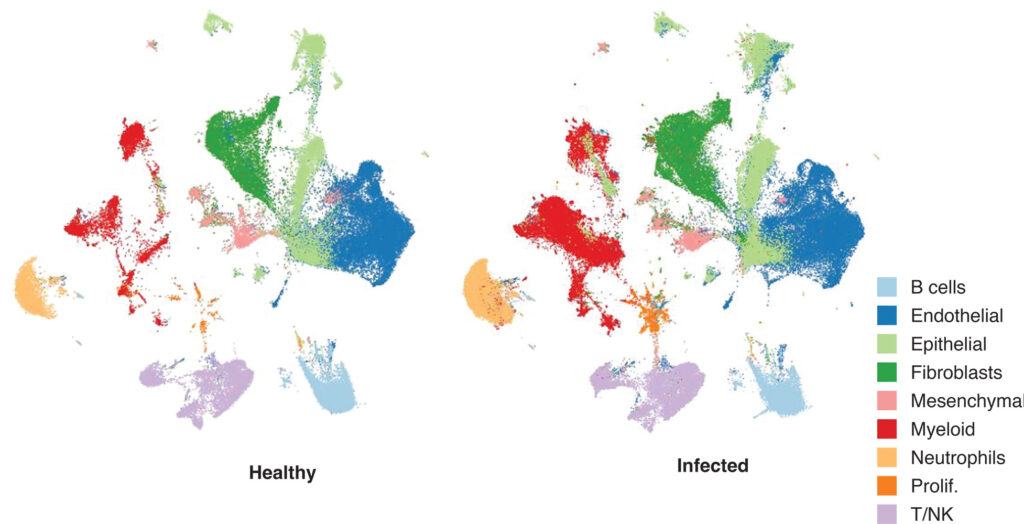
Experiments in which mice are exposed to a known pathogen under managed conditions (controlled pathogen exposure), enable researchers to study the development of an infection and the immune system response. By controlling the exact strain of the pathogen, the dosage levels, routes of exposure, timing, host factors, and environment, the team can establish potential cause and effect related to the immune system’s immediate response after infection, which cell populations respond first, how the disease progresses, and what leads to recovery or more severe illness. Another tool, scRNA-seq, measures gene expression in individual cells rather than averaging signals across an entire tissue sample, enabling researchers to identify which cells are inflammatory compared to which cells are behaving normally. In traditional bulk RNA sequencing, RNA extracted from thousands or millions of cells is pooled together before sequencing, obscuring differences among rare or functionally distinct cell populations. In contrast, scRNA-seq preserves cell-specific transcriptional information, enabling researchers to resolve how immune and nonimmune cells respond uniquely to infection and injury. To perform the analysis, lung tissue is first dissociated into a suspension of individual cells. Using a microfluidic droplet platform, single cells are encapsulated together with gel beads carrying unique molecular barcodes. Inside each droplet, messenger RNA released from the lysed (ruptured or broken) lung cell is captured and reverse-transcribed into complementary DNA (cDNA). During this process, each transcript receives both a cell-specific barcode and a unique molecular identifier (UMI), allowing researchers to distinguish where RNA molecules originated while also correcting for amplification bias introduced during polymerase chain reaction (PCR) amplification.
After barcoding and amplification, the team breaks the droplets and sequences the pooled cDNA libraries at high depth, generating hundreds of millions to billions of reads per experiment. Using computational pipelines to align the reads to the genome, Livermore’s scientists reconstruct transcriptomes for tens of thousands of individual cells simultaneously. The team uses graph-based clustering and dimensionality-reduction approaches such as uniform manifold approximation and projection (UMAP) to identify distinct cellular populations in infected and noninfected cells and visualize transcriptional relationships across experimental conditions.
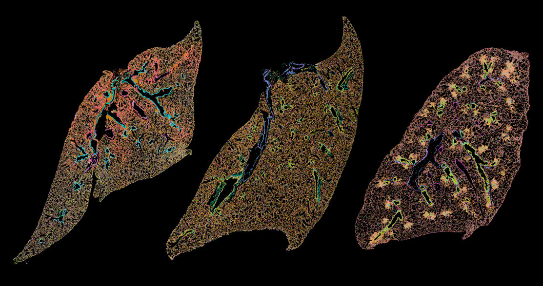
A third method the team applies is spatial transcriptomics, which measures gene activity in specific locations in the tissue. Cells do not act in isolation from each other, and in the case of infection, cancer, or tissue repair, location in the tissue is important. For example, a macrophage near a blood vessel could behave differently from one next to a damaged epithelium (densely packed cells that cover external body surfaces such as skin, line internal cavities and organs, and form glands). In infection studies, spatial transcriptomics, a visual representation of the location and clusters of different cell populations, can show where inflammatory cells accumulate, where antiviral or antimicrobial genes are activated, how immune cells organize around lesions and infected areas, how responses differ between damaged tissue and nearby healthy tissue, and how cell neighborhoods change over time. In the lung specifically, spatial transcriptomics can reveal inflammatory cells clustered around infected airways, repair programs in surrounding tissue, and distinguish immune zones in severe compared to mild diseases.
Exploring New Ground
Livermore’s biologists have conducted several individual studies on Valley fever (Coccidioides posadasii), Rift Valley fever virus, Venezuelan equine encephalitis virus, influenza, respiratory syncytial virus, bacterial pneumonia (Streptococcus pneumoniae), and COVID-19. “Our work didn’t start with the intent to generate an immune atlas,” says Sebastian, a Livermore staff scientist. “We were working on individual projects and asking specific questions. However, as we started to generate more data, we began to discuss standardizing the process of how and when we collect data and putting it into one big data set.” The immune atlas combines the data gathered from cells’ immune responses to the different diseases and then uses computational software to build visualization tools that map the varying responses across infected and noninfected cells.
Using scRNA-seq and spatial transcriptomics, the team sought to answer how the immune system responds inside an infected organ and why that response shifts from protective to damaging. “As we pulled together the data from millions of reads, we saw what cells were doing for tens of thousands of reads compared to other cells,” says Hum. “By putting the pictures together, we can tell the difference between a T cell and a normal epithelial cell, for example. And from this stage, once we have the resolution of what the cells are doing and how many there are, we can start comparing all the different time points, trajectories, and infection models.” Through several studies, the Livermore team found that severe disease is often driven not just by the pathogen itself but by specific immune programs that become harmful. The team’s methods helped identify which cells responded first (such as microglia in the brain and resident lung macrophages and stromal cells in the lungs), which cells entered later, where the cells went in the tissue, and whether they were helping or harming the individual.
To analyze this data and compare results across pathogens and different parts of the tissues being studied, Livermore’s team used a variety of computational tools. In each study, they collected data at multiple infection stages, looking for when changes occurred and when recovery signatures began to appear. Since every experiment was slightly different, the team integrated and visualized the data sets such that they could be superimposed over each other and compared. “As we quantify what cells enter and at what point in time, we can study how their profiles change,” says Sebastian. “For example, in a blood system, monocytes enter and differentiate into multiple subtypes. Some of these are highly correlated with better outcomes, while others are not. We want to identify what’s considered a good response compared to a bad response.” The comparisons indicated that the type of cells entering the lung in response to infection can influence the outcome.
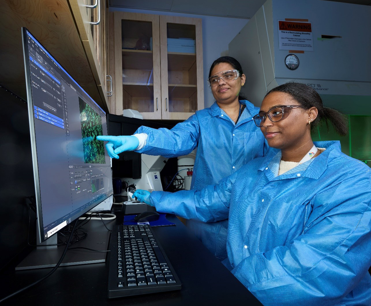
A series of computational tools have helped parse and present the data, including both R– and Python-based approaches for scRNA-seq, spatial transcriptomics, and multi-omic integration analyses. For scRNA-seq analyses, the team uses Seurat in R and Scanpy in Python, while spatial transcriptomic analyses are performed using Seurat in R and Squidpy in Python. Additional tools such as Scenic, Slingshot, and CellChat are used to identify gene regulatory patterns, examine changes in cell states and differentiation, and explore how cells may communicate with one another. Graph-based clustering methods were applied to define cellular populations, and UMAP is used for dimensionality reduction and visualization of cellular and molecular patterns across experimental conditions.
While still in the early stages of research, the team is already seeing interesting, sometimes unexpected, results. One macrophage population repeatedly shows up in multiple infection models, particularly lethal infections. These macrophages express many extracellular matrix proteins and matrix-remodeling enzymes that are more commonly associated with cell types such as fibroblasts. “We don’t know exactly what they do yet, but we can clearly see a correlation between some lethal infections and macrophages coming in and moving toward this kind of profile,” says Sebastian.
Charting a New Course
As the Laboratory looks toward future research opportunities, a key focus is to understand what patterns and results in mice can translate to humans. Weilhammer says, “Comparing infections in which the test subjects recover from the disease versus succumb to it is useful. In our mouse models of COVID, we saw young animals recover from infections while aged animals did not. However, that’s not the whole story—we’re also interested in how the genetic background of the host can impact the trajectory of the disease.” In the COVID-19 pandemic, even though younger people suffered less severe illness compared to more elderly people, this outcome was not universally the case—many young people also had severe infections, difficult recoveries, and fatalities. “We think it’s highly likely that there’s a strong genetic component behind why some people do well following infection and others do not,” continues Weilhammer.
In future studies, the team plans to integrate AI and deep learning models more significantly into their data analysis process and to help better predict an individual’s treatment needs and recovery path. “The basic research is difficult to pursue unless someone has all the experimental and computational capabilities that we have at Lawrence Livermore,” says Hum. “We’re still working toward the point where we can identify which illness someone has and how severe it might be from just a cough or other common symptom.”